Type 1 and Type 2 are two MRI patterns in the bone marrow beside a disc. Type 1 is dark on T1 and bright on T2, an oedema-like pattern. Type 2 is bright on T1, where fatty marrow has replaced normal marrow. A review did not establish that either type is linked to more pain. Modic · 1988 Herlin · 2018
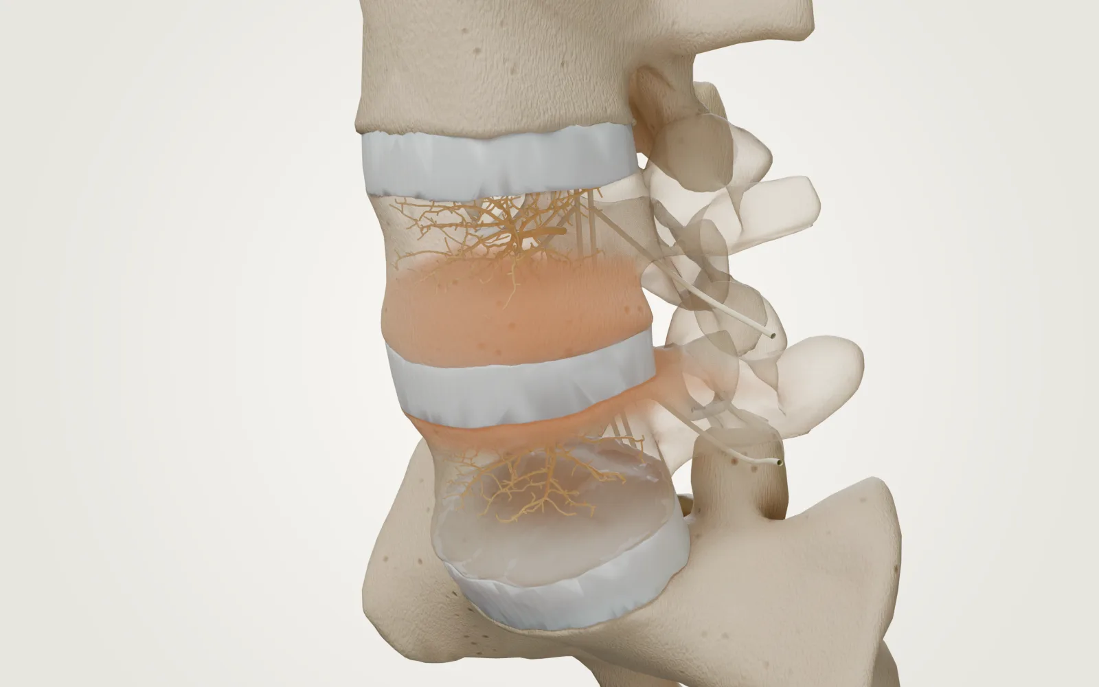
What is different?
| Type 1 | Type 2 | |
|---|---|---|
| Tissue pattern | Oedema-like change / vascularised fibrous tissue | Replacement with fatty marrow |
| T1 signal | Lower (darker) | Higher (brighter) |
| T2 signal | Higher (brighter) | Often higher or intermediate |
| Fat-suppressed sequence | Oedema signal can remain conspicuous | Fat signal is suppressed |
| What the label proves | A marrow imaging pattern | A marrow imaging pattern |
Is Type 1 more painful?
A scan label is not a pain ranking
Herlin · 2018Associations between Modic changes and back pain were inconsistent. The review did not establish a difference between Type 1 and Type 2.
- Who was studied
- Clinical and non-clinical populations across observational studies
- Compared with
- With versus without Modic changes
- Outcome & follow-up
- Association with pain and activity limitation · Cross-sectional and cohort evidence
- Limits
- Association is not causation; heterogeneous studies, mostly at risk of bias.
- Funding
- Research foundations and chiropractic research funding; see article disclosures.
A treatment study selecting people with a particular pattern does not make that pattern a diagnosis for everyone. Keep the question “what does the MRI show?” separate from “why does my back hurt?”
What about Type 3?
Type 3 describes a sclerotic pattern, with low signal on both T1 and T2. It is not shorthand for “the worst type”. Types can coexist. Modic · 1988
A useful next stepLook at the treatment evidence →