Modic changes are patterns seen in the marrow of a vertebral body, close to the endplate beside a disc. The term describes an imaging finding. It is not a complete diagnosis. Herlin · 2018
First, find the anatomy
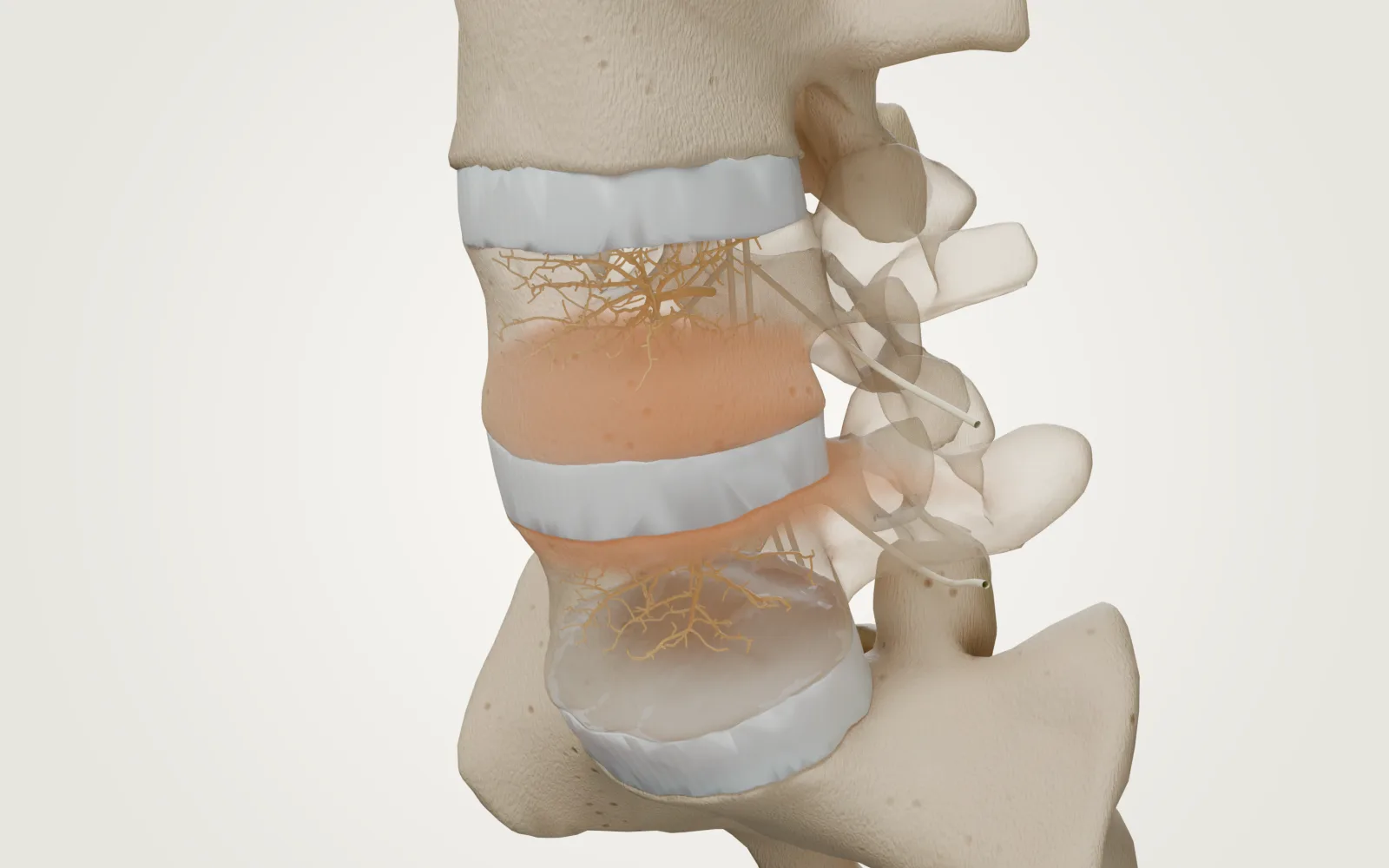
The shape is unchanged.
Modic describes a signal pattern on an MRI. Switching it on here leaves the same anatomy in place.
The change is in the marrow.
The coloured band sits beside the facing endplates. In Type 1, the signal is compatible with oedema-like and inflammatory change.
A different view of the same place.
Use the sagittal slice to look inside. The T1 and T2 schematics below show different signal contrasts. These are illustrations, not real scan images.
The cortexThe outer bone shell., marrowTissue within the vertebral body, beside the endplate., endplate, disc and nerve have different roles. “Endplate change” does not mean the same thing as a trapped nerve or a slipped disc.
What Type 1 looks like
Type 1 is low signal on T1-weighted images and high signal on T2-weighted images. Its tissue pattern is associated with oedema-like change and vascularised fibrous tissue. The warm colour in our model is an explanatory overlay, not the colour of an MRI. Modic · 1988
Three patterns, not three compulsory stages
| Pattern | Tissue description | T1 MRI | T2 MRI |
|---|---|---|---|
| Type 1 | Oedema-like / inflammatory pattern | Dark | Bright |
| Type 2 | Fatty marrow replacement | Bright | Often brighter or intermediate |
| Type 3 | Sclerotic, denser bone pattern | Dark | Dark |
These descriptions come from imaging and tissue studies. Mixed patterns can occur. A number is not a pain score, and people do not all move through the types in order. Modic · 1988
Does it explain the pain?
The pain question remains separate
Herlin · 2018Associations between Modic changes and back pain were inconsistent. The review did not establish a difference between Type 1 and Type 2.
- Who was studied
- Clinical and non-clinical populations across observational studies
- Compared with
- With versus without Modic changes
- Outcome & follow-up
- Association with pain and activity limitation · Cross-sectional and cohort evidence
- Limits
- Association is not causation; heterogeneous studies, mostly at risk of bias.
- Funding
- Research foundations and chiropractic research funding; see article disclosures.
A clinician considers your history, examination, other MRI findings and the broader pattern of symptoms. The scan alone cannot prove which structure is painful. Finding Type 1 should lead to a conversation, rather than an automatic treatment decision.
What happens over time?
Patterns may persist, mix or change. Repeat imaging cannot be turned into a reliable timetable for your symptoms. This guide does not use a claimed percentage for improvement within a year: that figure was not established in the primary evidence reviewed here.
Six questions to take with you.
You do not need to understand every technical term before your appointment.
- Where are the changes, and which MRI sequences show them?
- What makes you think they do, or do not, explain my pain?
- What other possible pain sources have you considered?
- What is a realistic plan for conservative care, and how will we review progress?
- For any proposed treatment, how closely do I match the people in its trials?
- What are the benefits, harms, alternatives, costs and reasons it might not help?